Dental X-rays, also known as radiographs, are essential diagnostic tools in dentistry. They allow dentists to see what’s happening beneath the surface of your teeth and gums, helping them identify and treat various oral health issues. In this comprehensive guide, we will explore the different types of dental X-rays commonly used in dental practice, along with their specific uses and benefits.
The Importance of Dental X-Rays
Dental X-rays provide valuable information that is not visible during a regular dental examination. They help dentists:
1. Detect Dental Issues Early: X-rays can reveal dental problems in their early stages, allowing for prompt treatment and prevention of complications.
2. Evaluate Tooth Decay: X-rays can detect cavities between teeth, which are not visible to the naked eye.
3. Monitor Tooth Development: In pediatric dentistry, X-rays help monitor the growth and development of children’s teeth.
4. Assess Tooth and Jaw Alignment: X-rays help orthodontists assess the alignment of teeth and jaws, aiding in the planning of orthodontic treatments.
5. Plan Dental Procedures: X-rays are crucial for planning procedures like tooth extractions, root canals, and dental implants.
6. Diagnose Gum Disease: X-rays can reveal the extent of gum disease and the condition of the jawbone.
Common Types of Dental X-Rays
1. Bitewing X-Rays:
Bitewing X-rays are the most common type of dental X-ray. They are named for the small, wing-like tabs on the X-ray film that patients bite down on during the procedure. Bitewing X-rays are used to detect cavities between teeth, evaluate the fit of dental fillings, and monitor bone density.
2. Periapical X-Rays:
Periapical X-rays focus on a single tooth from root to crown. They are used to assess the entire tooth structure, including the root and surrounding bone. Periapical X-rays are crucial for diagnosing dental issues like root infections, fractures, and abscesses.
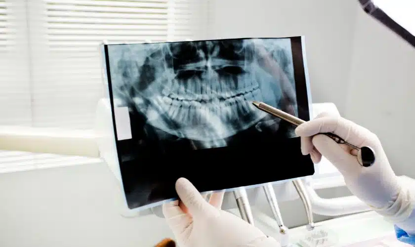
3. Panoramic X-Rays:
Panoramic X-rays provide a broad view of the entire mouth, including the teeth, jaws, and surrounding structures. They are particularly useful for orthodontic treatment planning, identifying impacted teeth, and assessing the temporomandibular joint (TMJ).
4. Occlusal X-Rays:
Occlusal X-rays capture the entire arch of teeth in one X-ray. They are typically used for young children who may have difficulty biting down on the small tabs of bitewing X-rays. Occlusal X-rays help detect dental issues in the early stages.
5. Cephalometric X-Rays:
Cephalometric X-rays focus on capturing the side profile of the face and skull. They are commonly used in orthodontics to assess facial growth, jaw alignment, and the positioning of teeth. Orthodontists use cephalometric X-rays to develop personalized treatment plans.
6. Cone Beam Computed Tomography (CBCT):
CBCT is a three-dimensional imaging technique that provides detailed, cross-sectional images of the teeth and jaws. It is invaluable for planning complex dental procedures like dental implant placement, orthodontic treatment, and oral surgery.
Radiation Safety in Dental X-Rays
While dental X-rays are essential for diagnosis and treatment planning, safety is a paramount concern. Dentists follow strict guidelines to minimize radiation exposure. Here are some safety measures:
– Lead aprons and thyroid collars: Patients are provided with lead aprons to shield the body from radiation. Thyroid collars protect the thyroid gland.
– Digital X-rays: Digital technology reduces radiation exposure by up to 80% compared to traditional film X-rays.
– As Low As Reasonably Achievable (ALARA) principle: Dentists adhere to this principle to keep radiation doses as low as possible while obtaining the necessary diagnostic information.
Frequency of Dental X-Rays
The frequency of dental X-rays varies depending on a patient’s age, oral health, and risk factors. Generally, the following guidelines are followed:
– Bitewing X-rays are typically taken annually for adults to check for cavities.
– Periapical X-rays are taken as needed for specific dental concerns.
– Panoramic X-rays may be taken every 3 to 5 years for adults.
– Children and adolescents may require more frequent X-rays to monitor growth and development.
Dental X-rays are indispensable tools in modern dentistry, allowing for accurate diagnosis and effective treatment planning. By understanding the different types of dental X-rays and their specific uses, patients can appreciate their role in maintaining optimal oral health. Dentists prioritize patient safety by following stringent guidelines to minimize radiation exposure, ensuring that X-rays are a valuable and safe aspect of dental care. Regular dental check-ups and X-rays, as recommended by your dentist, are key to maintaining a healthy and beautiful smile throughout your life.